Q: I have to undergo colon cancer surgery. Does that mean my colon will be removed? How do you do the surgery?
A: Just a quick refresher: The large intestine, or colon, is the final part of the digestive tract and its job is to absorb water back into the body and create waste. The colon is an average length of about 1.5 metres with the final 15 cm being the rectum and then the final sphincter muscle that controls the stool is the anus.
To answer your question in a nutshell, usually most of the colon is left alone and only the section with the cancer is removed, and the two ends are brought together to create a connection. Imagine it like cutting out a damaged part of a garden hose and reattaching the two ends.
Let’s explore the details of this kind of surgery more below.
Location, location, location
How the colorectal cancer surgery is done is essentially based on location. At the time of the colonoscopy, the colorectal cancer is diagnosed and the exact location is the cancer is identified. This is the critical information the surgeon will use to plan the surgery. There are lots of other factors that determine how much colon should be removed to treat the cancer.
Distance: The most important consideration is the safe distance from the cancer to make absolutely sure that there are no cancer cells left behind; this is called the margin. Studies have shown that in colon cancer, the resection lines should be about 10 cm away from the cancer on both sides. In rectal cancer, if the cancer is low in the rectum and very close to the anus, we accept as little as a 1 cm margin if it means that we can reconstruct the intestine and not remove the anus. If the cancer is at the sphincter muscle and involving the anus or very close to it, than the entire anus must be removed and in those cases a permanent stoma is necessary (which I’ll describe in more detail in another post.)
Lymph Nodes: The colorectal cancer resection also must include all the lymph nodes or glands that surround the tumour. A pathologist examines the removed nodes after the surgery to see if cancer cells had travelled to the nodes. This helps us decide which patients require chemotherapy after the surgery.
Anatomical considerations: There are other things we look at in order to plan the operation, and this is determined by understanding the blood flow to the colon. After a segment of the colon is removed, it is extremely important that the two ends that are used to make the new connection have a very good blood supply in order to make sure they heal to each other and there is no leak from the connection. To be sure the blood supply to the two ends of colon is ideal, there are very defined sections of the colon we remove and we are very particular about the location of the two ends that we bring together. Another critical anatomic consideration that the surgeon must be certain of is that the two ends that are brought together to reconnect are under absolutely no tension whatsoever, as this also may lead to a leak in the connection.
Based on all these factors, the surgeon will decide what segment of the large intestine they will need to remove in order to:
- Be absolutely certain the cancer is completely removed with all its associated nodes.
- Optimize the chances of healthy well-healed reconnection of the two ends.
Therefore the surgery can entail removal of the right or left side of the colon, sigmoid colon, upper rectum, entire rectum, or even the entire rectum and anus. These surgeries are all described here.
Open surgery or laparoscopic?
The surgeries for colorectal cancer removal can be done both open and laparoscopically. Open surgery involves a large vertical incision in the middle of the abdomen and the operation is done through this cut. Laparoscopic surgery involves placing a tiny camera in the belly button (umbilicus) and three tiny incisions in the abdomen. We use long surgical instruments, and do the operation by looking in the abdomen via the camera on a large screen.
When possible and safe, and in the right setting, the laparoscopic approach is, quite frankly, better. Much less pain, shorter recovery, shorter time in hospital and no wound issues. Many large trials have shown that the cancer results are the same for open and laparoscopic surgery, as long as the surgeon is comfortable with that approach and has ample experience and volume of cases. My personal opinion is that laparoscopic surgery for colon cancer should be the standard of care. Having said that, many cancers are not safe to remove laparoscopically because they are too large, or they are stuck to other organs, or the patient has had many other surgeries and there is too much scar tissue. But the laparoscopic approach should always be considered and discussed.
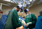
Inside my OR
In my practice about 90 per cent of the surgeries are done laparoscopically so I’ll explain that in a bit more detail:
Step 1 – Insufflation: we insert a small metal tube into the belly button and through it we blow air into the abdomen. This air gives us a working space by separating the abdominal wall from all the organs. We then insert the camera into this space.
Step 2 – Surgical Access: we insert three more small metal tubes into the abdomen. We place our very long, thin surgical instruments through these tubes. We control the instruments from outside the abdomen but they function inside the abdomen. We can see everything in the abdomen through the camera.
Step 3 – Decision Making: we identify the exact area that needs to be removed by looking at the cancer and then considering all the factors mentioned above in order to decide on exactly where we will cut.
Step 4 – Resection: using a tiny mechanical stapler that slides into the metal tubes, we cut both ends of the colon. The stapling device seals the colon closed before cutting, so there is no contamination. We ensure when we remove it that we’ve included all the lymph nodes surrounding it.
Step 5 – Reconstruction: we first cut away any scar tissue to loosen the two ends so they will come together easily now that a segment has been removed. Then with a combination of stapling and sewing, we make a new connection of the two ends of colon.
Step 6 – Extraction: we make a small cut through the skin to remove the diseased segment of the colon from the abdomen and this ends the procedure.
This step-by-step list makes it all sound very straightforward — and it certainly can be for early small cancers in favourable locations. But it can also be very complex and require much more involved surgeries to get the cancer out and create the best possible chance to cure the colorectal cancer. In very rare occasions for cancers that are too close to the end to make a connection, a permanent pouch for stool can be required.
If you have questions about your specific surgery, please feel comfortable to talk to your surgeon. Ask questions until you understand. It is your body, and you have the right to understand what the surgery will entail.