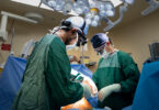
The MOLLI team from left: Mark Semple, Alexandru Nicolae, Dr. Nicole Look Hong, Ananth Ravi, John Dillon.
(Photography by Kevin Van Paassen)
A routine mammogram detected a suspicious mass in Sieu-Mui Luc Ong’s right breast. An ultrasound and biopsy confirmed it: the 74-year-old had breast cancer.
“The tumour was found early and a lumpectomy was recommended,” says Sieu-Mui, who lives in Thornhill, Ont. “I felt comfortable with that and I was not too afraid.”
The clinical trials team approached Sieu-Mui about MOLLI (magnetic occult lesion localization and imaging), a new tool that could help the surgeons locate and remove the tumour in her breast with ease.
Think of MOLLI – a device designed and made in Sunnybrook – like a stud finder, the handheld metal detector used to locate studs behind drywall. A tiny magnet is inserted into the breast tumour and, during surgery, the surgeon uses a small wand to locate the magnet. The system, to which the wand is connected, then identifies the location and exact depth of the magnet, showing the surgeon where and how deep to cut.
Sieu-Mui became the first patient in a pilot trial testing the effectiveness of the device. “I don’t mind being first,” Sieu-Mui says. “The researchers and doctors have to test it out, and then it can help people in the future.”
During a lumpectomy, surgeons try to remove the tumour with clear margins, so no cancer cells are left behind.
“As surgeons, we don’t have X-ray vision, so we need something to guide us to the tumour and help us remove it as precisely as possible,” says Dr. Nicole Look Hong, a breast cancer surgeon at Sunnybrook and lead physician on the MOLLI project. “If, after lumpectomy, it’s found [that] the margins aren’t clear of cancer cells, the patient has to come back for more surgery.”
Guide wires or, more recently, radioactive seeds implanted into the tumour help surgeons locate where to make the incision.
But the guide wires are painful for patients, and the radioactive locator seeds (not to be confused with radioactive seeds used to deliver radiation therapy for other types of cancer) have a host of administrative costs and processes that make them impossible for some cancer programs to adopt.
The radioactive seed system also doesn’t tell a surgeon how deep to cut. That means there’s an element of educated guessing, Dr. Look Hong points out.
The topic of lumpectomy navigation and radioactivity came up at a summer barbecue party about seven years ago at the home of Dr. Calvin Law, chief of the Odette Cancer Centre.
Dr. Look Hong connected with medical physicist Ananth Ravi and they set out to find a better, cheaper way to locate tumours.


Gold markers
It has taken Ananth, Dr. Look Hong and a team of engineers lots of time, testing and trips to the hardware store, literally, to get MOLLI to this point.
“The first step we take when trying to solve a problem is identifying all the possible solutions, then choosing ones that could work for the health-care system,” says Ananth. “One of our first attempts was to buy a stud finder and use it to try to find implantable gold markers.”
The stud finder with gold worked, but only if the gold wasn’t too deep, he notes. They “souped up” the device to try detecting the gold deeper in tissue. “It became too powerful and it blew up,” he says.
The team played with a few other ideas like copper tubing and infrared goggles (too hot) and microchips (too big) before returning to the stud-finder concept.
“In health care, the solution you come up with has to work all of the time,” Ananth explains. “The simpler it is, the less chance there is of it breaking.”
His team turned to very weak magnets and, using their stud-finder tests as inspiration, set out with the surgeons to design and create a wand and computer interface. The wand is 3-D-printed in medical-grade materials, and the custom software was all designed and made in-house, too.
“It’s so detailed and precise but also so simple,” Dr. Look Hong says. “Instead of getting more complicated, the concept has gotten simpler – not to underestimate the years of work that have gone into it.”
Both Dr. Look Hong and Ananth are pleased with the result – and ecstatic the system is being tested in patients.
“This gives us clear measurements,” Dr. Look Hong says. “Because the magnetic seed doesn’t decay like the radioactive seeds, it has a constant signal, so we can get a measurement from the seed to the wand. If I know reliably that I’m two centimetres away from the tumour, then I know it is a safe place to cut to have good margins.”
What’s next?
According to Sieu-Mui, when the magnet was implanted in her tumour, she barely even noticed it. She had both the radioactive seed and the new MOLLI seed put in, so that the team could compare the two systems. She returned two days later to Sunnybrook for the lumpectomy under anaesthetic and went home the same day. There’s hardly even a mark on her breast, she says.
Dr. Look Hong hopes the MOLLI system will rise from its humble beginnings to become a global standard of care.
“MOLLI – a magnet, a small wand and a screen – would be easy for more centres to incorporate, even in low-and middle-income countries where they don’t screen for breast cancer because the only treatment they have available is full mastectomy,” says Dr. Look Hong. “The goal is that MOLLI can be disseminated widely at a low cost and help save lives.”