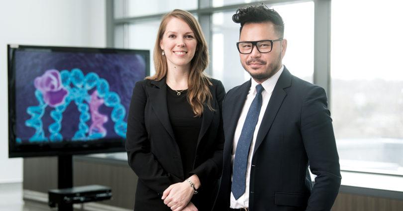
Dr. Katarzyna Jerzak and Dr. William Tran. (Photo by Kevin Van Paassen)
One-size-fits-all has no place in Sunnybrook’s personalized breast cancer program. Using imaging, innovative genomic approaches and high-tech cellular analysis, doctors and scientists are taking a bold new path to treat breast cancer with precision-based medicine.
Debbie Duclos had a gut feeling something was very wrong with her left breast. It was November 2014 and the 42-year-old registered nurse from Campbellville, Ont., noticed that its shape and feel had changed.
After a local hospital ordered a mammogram and an ultrasound, “a sizeable area of concern” was identified through imaging. Stricken, Debbie conferred with her family doctor and was referred to Sunnybrook for her biopsy, having worked at the hospital in the past.
“Within 48 hours, I had my diagnosis. Stage 2 breast cancer,” she recalls. “The tumour was the size of a lemon.”
The news led to a lot of anxiety and fear.
“The uncertainty of a cancer diagnosis really rocks your boat,” says Debbie. “I was thinking, ‘Why me? Why did this happen?’”
Reeling from the news, Debbie was comforted by the support of her husband, parents and friends, and by the fact that she had an appointment with a team of oncologists at Sunnybrook by the end of the week.
During that first meeting, the team of doctors and radiologists provided Debbie with a detailed plan for her care. Within the next nine months, they were going to give her chemotherapy, surgery and radiation, while continuously tracking her tumour’s response to treatment.
She was also enrolled in several studies during her breast cancer journey, including a tumour-mapping study.
Debbie left the meeting understanding her diagnosis and with a personalized plan, giving her an enormous feeling of relief.
“I felt like they had taken me under their wing,” she says. “They had a clear direction for me. It’s what enabled me to really think I was going to be okay.”
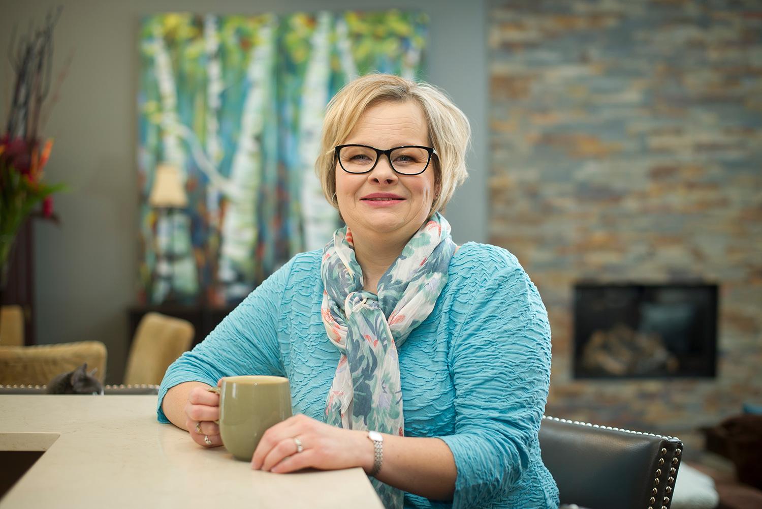
Debbie Duclos, diagnosed with breast cancer in 2014, benefited from Sunnybrook’s innovative personalized treatment program. (Photograph by Kevin Van Paassen)
A personalized approach
Debbie had become part of Sunnybrook’s ongoing clinical research program that aims to provide a personalized approach to cancer care.
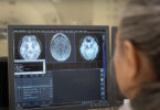
This approach focuses on obtaining as much information as possible about tumours through quantitative magnetic resonance imaging (MRI), digital pathology and genetic mapping, known as genomics. Using these advanced techniques, Sunnybrook oncologists and scientists work together to determine exactly how these tumours behave – and what treatments are best at eradicating them.
“We decided to form a core team of clinical and scientific experts here at Sunnybrook,” says Dr. William Tran, a radiation therapist and clinician-scientist at Sunnybrook and a vital part of the new precision medicine program. The team includes Dr. Katarzyna Jerzak, a medical oncologist at the Odette Cancer Centre and assistant professor in the Department of Medicine at the University of Toronto, pathologists and radiation oncologists at Sunnybrook and Dr. David Andrews, director and senior scientist in the Biological Sciences Platform at Sunnybrook Research Institute and professor of biochemistry at the University of Toronto.
“Now I can answer patients who ask, ‘How do you know if the chemo is working?’”
– Dr. Katarzyna Jerzak,
medical oncologist at the Odette Cancer Centre at Sunnybrook
They work holistically with patients at every stage of their care program to assess the responsiveness of the tumour to the treatment.
Traditionally, tumours aren’t viewed multiple times over an extended period during treatment, says Dr. Tran. The new, personalized approach tracks the cancer as it evolves in a patient’s body. In addition, the information gleaned from these patients will be tracked, recorded and stored in a databank, to be used for guidance on future cases at Sunnybrook and other Canadian hospitals.
Dr. Tran says there is a great interest in the medical and scientific community to improve treatments for breast cancer. “We’re trying to shake [things] up in this research program.”
‘A changing paradigm’
As part of the precision-treatment program, the team is trying to identify women who are at high-risk for developing metastasis (the spread of the disease beyond the primary site). Once a patient with high-risk breast cancer is identified by medical oncologists at Sunnybrook, they are followed by the team as part of a study.
At the onset of treatment, these patients receive chemotherapy to shrink their tumours. This approach can halt the cancer cells early and help prevent metastases, even prior to surgery.
“There’s a changing paradigm now,” says Dr. Jerzak. “Historically, women with early breast cancer were treated with surgery first, followed by chemotherapy. But increasingly, women with triple-negative or HER2 positive tumours are getting chemotherapy first, instead of surgery first.”
For some women, the upfront chemo means the tumour shrinks entirely or enough that breast conserving surgery is possible, rather than a full mastectomy.
At select points in the chemotherapy regimen, quantitative imaging will be done to determine if the tumour is responding well to treatment, says Dr. Tran.
“We are exploring new ways to analyze tumours using quantitative MRI, ultrasound and digital pathology; we think these are our best shot right now,” he says. Unlike conventional MRI, quantitative MRI (and imaging) provides clinicians with measurable and consistent data of the biological and physiological properties of the tumour.
More good news
In accordance with the Sunnybrook team’s personalized approach, Debbie underwent chemo immediately after her diagnosis. She watched as her tumour shrank dramatically.
“[The doctors] could see how the chemo was reducing the tumour size,” Debbie recalls. “And they were able to give me results.”
She later had a mastectomy of her left breast, 36 lymph nodes removed and 25 sessions of radiation.
Dr. Jerzak is happy that she now has more good news to share with patients like Debbie.
“Now I can answer patients who ask, ‘How do you know if the chemo is working?’” she says.
Dr. Jerzak says she’s currently working to recruit patients with triple-negative or HER2 positive breast cancer for a study that not only incorporates imaging, but also measures blood-based and genomic markers of response to
chemo-therapy. Later, she hopes to work with women with metastatic breast cancer as well.
Databank for the future
A biopsy of a tumour taken at the onset of treatment can provide scientists with vital information about its composition. Tumours are often made up of a variety of cancer cells – rather than just one type – meaning that each portion of a tumour can react differently to different medications.
That’s why after Dr. Andrews and his team remove a tumour sample, they culture the cancerous cells in the lab. Then, they effectively grow 3-D models of the tumours in order to determine their composition and their responsiveness to a variety of drugs. (See below.)
“We try to extract a lot of information from images of the cells,” says Dr. Andrews. “We’re trying to find out how they’ll respond to treatment, [and] within weeks, instead of months, we have an answer.”
The end game, says Dr. Andrews, is that the patient will be given targeted chemotherapy to treat the exact cancer – or cancers – they have, rather than being bombarded with drugs that wipe out not only cancer cells but healthy cells as well.
And the work done in the lab won’t just benefit each individual patient. All the information being gathered from patients is being stored, with the hopes of building a large databank in the future that doctors can access.
In essence, the databank will illustrate what worked – and what didn’t – for each patient and their particular type of tumour. This valuable information will be used to help oncologists plot the best treatment plans for future patients.
Giving back
Now 46, Debbie has been in remission since September 2015. She gets follow-up appointments every six months and is still undergoing active surveillance as part of the precision medicine studies. Last year, imaging caught some shadowing in her right breast, which could indicate a tumour. Luckily, the area hasn’t changed in subsequent scans, and Debbie is confident she’s being watched carefully by her team at Sunnybrook.
She is now the founder and CEO of a medical-device training company, having been inspired by some of the technology used during her care.
Debbie’s also been volunteering in her community, including participating in events supporting women’s cancer research. She says she’s happy to share her energy and enthusiasm, having received a new lease on life.
“I am just so grateful to my entire care team and want to show my appreciation by giving back,” she says. “I always say, ‘Is there a way you can pay it forward?’”
Solving complex cases
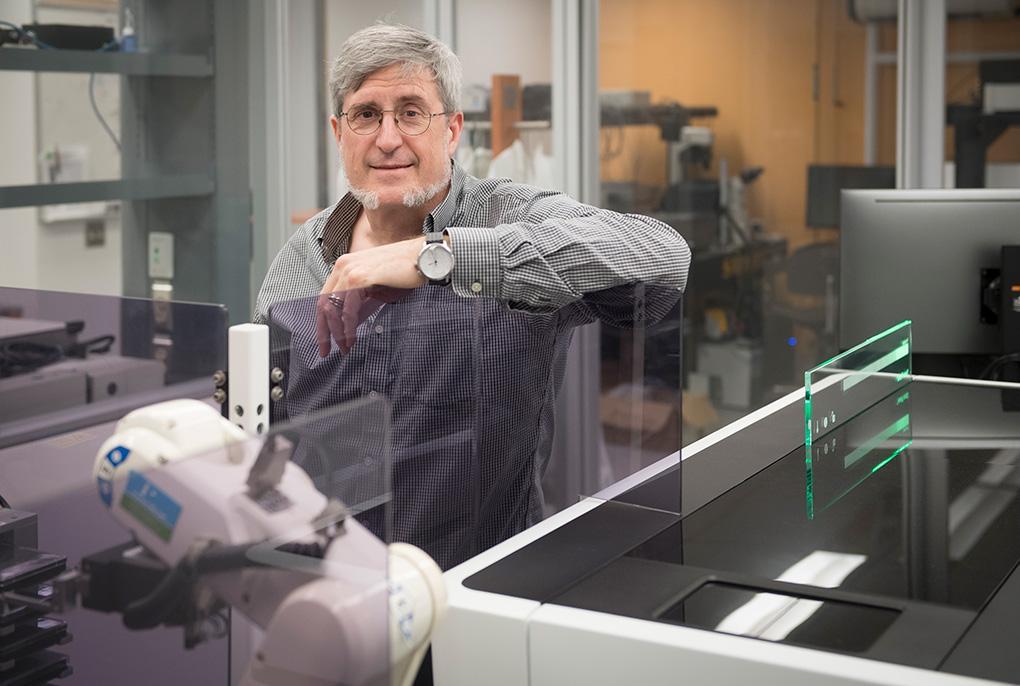
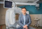
Dr. David Andrews in Sunnybrook’s High Content Screening Lab. (Photograph by Kevin Van Paassen)
In the High Content Screening Lab, at the Sunnybrook Research Institute, senior scientist Dr. David Andrews and his team are growing cancerous tumour cells sourced from breast biopsies into 3-D models called organoids. These models can provide key information about the cells within them – and give clues as to which medication combinations will prove successful.
“We’re trying to mimic what’s going on in the patient’s body,” explains Dr. Andrews.
Zeroing in on the ‘bad guys’
Some of his research looks at how cancer driver cells work. These cells, which are responsible for fuelling tumour growth, are notoriously tough to treat. (“They’re the real bad guys,” he says.) Dr. Andrews wants chemotherapy to be more precise, zeroing in on these types of cells that resist chemotherapy and often come back.
To that end, he and his team analyze how the cancer cells in his samples behave. These behaviours can offer clues as to how effective chemotherapy will be. “In response to chemotherapy drugs, we ask, ‘Are they stressed? Are they starving? Are they going to sit there and wait it out? Or are they going to die?’ Chemotherapy regimens can evoke those behaviours,” says Dr. Andrews.
Ultimately, scientists won’t just be analyzing cellular behaviour. Dr. Andrews says scientists may someday be able to activate a built-in self-destruct program in these tough-to-treat cancer cells, instructing them to die off.
Predicting the right treatment
To witness this kind of sophisticated cellular interplay, Dr. Andrews and his team use state-of-the-art technology. New, automated microscopes generate data sets containing millions of images. This data is then evaluated though a process called high content analysis – using microscopes and computers to employ complex algorithms that can identify and categorize cells.
Once the information on the cells is available, it is then stored in the lab’s servers. The information can then be accessed by radiologists and oncologists with the goal of helping them treat high-risk breast cancer patients.
“The goal is to be able to tell patients what drug combination they should be taking,” says Dr. Andrews. “We need a way to predict success upfront.”