This month, the city’s first 7-Tesla (7T) magnetic resonance imaging (MRI) machine was delivered to Sunnybrook. The machine, which is now housed next to the new Garry Hurvitz Brain Sciences Centre, is a powerful scanner, producing images in unprecedented detail, allowing researchers to see pathologies in the nervous system never seen before with MRI.
The 7T MRI scanner is part of the Toronto Neuro-Immunology/Imaging Consortium (TONIIC), a multi-site collaborative research initiative focused on neuroimmunology and neuroimaging for diseases such as stroke, multiple sclerosis and cancer. TONIIC is made up of hospital and academic institutions across Toronto, including Sunnybrook, Baycrest, SickKids, University Health Network (UHN), Centre for Addiction and Mental Health (CAMH), Unity Health Toronto (St. Michael’s Hospital) and the University of Toronto’s Temerty Faculty of Medicine. Although construction of the facility is still underway, 7T MRI will soon transform the way Toronto researchers see, understand and study novel approaches for diagnosing, monitoring and treating the brain.
The scanner, manufactured by Siemens Healthineers, is one of only three clinical 7T systems in Canada. Getting the scanner here from Europe was a multi-stage effort. It was flown to Chicago on one of the few heavy-lift planes equipped to transport the 7T magnet. From there, it was transported by road the rest of the way to Toronto. The 7T MRI scanner was maintained on route with liquid helium to keep its inner core extremely cold. This minimized the amount of additional liquid helium that will be needed to fill the magnet in Toronto during installation, when electric current will be applied to achieve the ultra-high magnetic field strength of 7T.

The 7T magnet arriving at Sunnybrook Health Sciences Centre.
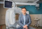
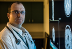
Dr. Kâmil Uludağ and Dr. Simon Graham, senior scientists in the Physical Sciences and Hurvitz Brain Sciences Program at Sunnybrook, are both harnessing 7T MRI technology to explore fundamental methodological research and to advance clinical applications of MRI. The focus of Dr. Uludağ’s work is integrating MRI techniques into clinical research for conditions such as brain tumors, depression, OCD, neurodegeneration and Alzheimer’s disease. Dr. Graham’s work focuses on the applications of MRI of brain activity in healthy individuals as well as patients suffering from stroke, Alzheimer’s Disease, brain cancer and traumatic brain injury, as well as improving the use of MRI and other associated medical devices.
We spoke with them to learn more about the power of 7T MRI and its potential to revolutionize research, care and outcomes for patients living with brain conditions.
How does an MRI work?
An MRI scanner is a type of diagnostic tool crucial for informing the diagnosis and treatment of many diseases, including brain conditions such as cancer, dementia, Parkinson’s disease, multiple sclerosis (MS) and amyotrophic lateral sclerosis (ALS).
When patients enter an MRI scanner, they’re exposed to a strong magnetic field that causes the magnetic hydrogen nuclei of water molecules in the body to align in one direction — like the earth’s magnetic field moves a compass needle. Electromagnetic waves are then sent through the body, causing the magnetism of the hydrogen nuclei to swing out of alignment, and then realign again. This reaction, which can be slightly different for healthy and diseased tissues in the body, is recorded and an image is created.
How is the 7T MRI different from other MRIs?
There are different strengths of MRIs, which are measured in Tesla (T) — the unit of measurement representing the strength of the magnetic field, which is usually set at either 1.5T or 3T for hospital MRI scanners.
The 7T MRI has a higher magnetic field than the 3T or 1.5T MRI, which permits higher resolution imaging. This results in a more detailed and less grainy image, enabling scientists and clinicians to see previously invisible features of the brain.

Many brain conditions like cancer or dementia, start in extremely small parts of the brain and later grow and expand to other parts of the brain and body. Many brain conditions may also appear similar on MRI scans performed at lower field strengths, making diagnosis difficult. With higher resolution and more detailed images, clinicians will have the ability to detect disease earlier, and with greater accuracy. This will enable existing treatments, and new treatments under development, to be applied earlier towards improved patient outcomes.
What are some use cases for the 7T MRI?
The new 7T MRI will be used to help researchers advance the way many brain disorders are detected, diagnosed and monitored. It has the potential to support timelier diagnosis of conditions that appear small in the brain but have a big impact on an individual’s health, like brain lesions or microbleeds. The higher field strength produced at the 7T will make these conditions more visible on scans.
Researchers will also be using the 7T to help support the development of precision treatments, such as more precise targeting in MR-guided focused ultrasound, deep brain stimulation or improved radiation therapy treatment for patients with cancer. Cancer tissue can be hard to distinguish from other types of pathologies after radiotherapy and the 7T can provide more insight on how a patient’s cancer reacts to the treatment.
In other research, 7T MRI will be used to monitor treatments and disease progression. The images will give scientists a more detailed look at how new procedures and drugs impact the progression of different neurodegenerative conditions like Alzheimer’s or Parkinson’s disease.
How will the 7T MRI advance patient care and outcomes in the city?

Moving the 7T magnet into its new facility next to the Garry Hurvitz Brain Sciences Centre.
The ultra-high magnetic strength of 7T MRI will result in images of the brain with more detail than ever before, enabling researchers to get a closer look and better understanding of how disease impacts the brain, ultimately supporting earlier detection and diagnosis of disease and more personalized and precise treatments.
The new 7T will have a significant impact on patients across the entire city. Although the scanner is located at Sunnybrook, as part of the TONIIC research network, it will support research and advance discoveries in medical imaging of the brain throughout Toronto.
Support for the 7T MRI scanner and TONIIC is provided by the Canada Foundation for Innovation (CFI) and the Ontario Research Fund.