It is sudden. It strikes without warning. And it is often fatal. Sudden cardiac death, often caused by a condition called ventricular tachycardia (VT), kills about 40,000 Canadians each year. Scientists at Sunnybrook Research Institute (SRI) are hard at work advancing imaging techniques to identify and reverse this ticking time bomb.
When a person has ventricular tachycardia, faulty electrical signals in the ventricles of the heart cause their heart to beat too fast or irregularly, impeding proper blood flow to the body. “This is a very urgent problem,” says Dr. Fumin Guo, a postdoctoral fellow working in the cardiovascular imaging lab of Dr. Graham Wright at SRI. “Most of these events occur, without previous symptoms, at home or in a public space, not in hospital. And they can be fatal within minutes. We are trying to improve diagnosis and therapy to prevent sudden cardiac death.”
One of the current treatments for ventricular tachycardia is radiofrequency ablation, which involves guiding a device into the heart and using an electrical current to heat up and destroy a small area of tissue that may lead to the abnormal electrical signals (arrhythmia). But about 35 per cent of ablation procedures result in either initial failure or later recurrence.
Preclinical work by Dr. Guo and others in Dr. Wright’s lab is focused on improving the outlook for those with VT in three ways: identifying the underlying structural and functional issues in the hearts of individuals, pinpointing damaged tissue with greater precision, and delivering treatment with more accuracy and efficiency.
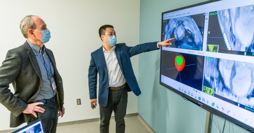
The lab has demonstrated success using 3D magnetic resonance imaging (MRI) to guide radiofrequency ablations with improved precision. “Imaging, and in particular MRI, shows great promise in helping to improve management in this patient population, and we are working at the state of the art to look at all aspects from identifying those at risk, to improving the effectiveness of procedures,” says Dr. Wright, who is a senior scientist in the Physical Sciences Platform and Schulich Heart Research Program at SRI.
Dr. Guo recently won the prestigious Polanyi Prize in Physiology and Medicine for his significant contribution to this work. With his background in biomedical engineering, he is developing, using artificial intelligence and computer vision methods, automated image analysis systems that map the heart and pinpoint the damaged tissue. He is also advancing computer algorithms to plan, guide and assess VT treatment.
“Fumin’s work means the image analysis needed to guide procedures will be more repeatable, faster, and hopefully more accessible to others,” says Dr. Wright. “The Polanyi Prize is very prestigious. Winning the prize is emblematic of the clinical relevance of the work and the excellence of Sunnybrook’s program in image-guided, personalized, precision therapy.”
Dr. Wright adds that his lab is collaborating with clinical heart programs and partnering with industry to ensure the preclinical work can be translated to patient care as quickly as possible.
For his part, Dr. Guo is enthusiastic about what lies ahead. “Our ultimate objective is to cure patients with ventricular tachycardia conditions. We want patients to live longer, healthier and happier.”


