It’s an emotional process that affects patients every day – after screening or surgery, they wait to hear if they have cancer.
Specimens, or tissue samples, are taken for further testing. So what happens to that specimen behind the scenes? And who are the experts at Sunnybrook tasked with determining what microscopic discoveries will mean for a patient moving forward?
The Department of Anatomic Pathology is where it all happens.
It’s comprised of a multidisciplinary team of 19 pathologists – medical doctors trained to make diagnoses based on microscopic examination of tissue – alongside 50 technical and administrative staff, including medical laboratory technologists and assistants, pathologist assistants, clerical staff and management. Every year, this team transforms 30,000 surgical and 20,000 cytology (cell) specimens into glass slides that are scrutinized, one by one, by experts trained in various cancers.
“This is not just a lab,” notes Dr. Nadia Ismiil, chief of anatomic pathology. “Our highly trained staff give diagnostic opinions that will help guide treatment [for patients] and their future. Every report is unique and individualized, and we take great pride in being an important part of each patient’s journey.”
Here’s a closer look at the steps each specimen undergoes.
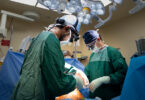
Each patient’s specimen is logged into the laboratory information system. The tissues are then fixed in formalin – a colourless solution of formaldehyde in water – and examined visually by pathologist assistants.

Abnormal-appearing areas are selected for further processing.

These areas of tissue are embedded in paraffin wax…

so they may be cut into four-micrometre-thick sections…

… mounted on glass slides and stained by medical laboratory technologists. The staining process takes about one hour and helps the cells to be seen more clearly.

The mounted, stained tissue is then examined under a microscope (E) by pathologists. Several sections from each sample need to be examined at various depths, when looking for possible cancer cells. Pathologists may request additional studies to be performed on the tissue when needed. With all this information, the pathologist can then issue a personalized report on a patient’s cancer.
(Photographs by Doug Nicholson)